DR. VINAY MAHENDRA
Kidney Stone Robotic Surgeon in Kolkata, India
MS. Mch (Urol.), DBN (Urol.), FRCS (Glasg.) FRCS (Urol.) Senior Consultant Urologist
Laparoscopic And Robotic Urological Surgeon and Renal Transplant Surgeon
![]()
FOR APPOINTMENT CALL SECRETARY
+91 9830216715 (Secretary Amit Roychowdhury : 11:00 A.M. - 4:00 P.M.)
+91 9830492572 (Secretary, Dalia Chatterjee : 11:00 A.M. - 4:00 P.M.)
STONE ROBOTIC SURGERY
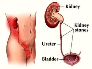
Kidney stones are very common, they occur in approximately 12% of people.
They are very common and unfortunately, extremely painful.
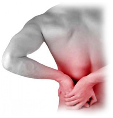
Patients usually present with pain in the loin which radiates down to the groin area and they often have nausea and vomiting.
Itís important to check that they donít have a fever. One cause of fever could be an obstructed and infected kidney. This is a medical emergency and we have to drain that kidney straight away.

Our management generally depends on initially diagnosing the problem.
So the way we diagnose the problem is that we do
• A urine test: we check if thereís blood in the urine. That gives us an indication that there might be a kidney stone. This also allows us to check that thereís no infection.
• Imaging tests: The imaging test that we do these days is a CT scan, a non-contrast spiral CT scan and this is very, very accurate and it usually tells us where the stone is, the size of the stone and the degree of blockage or obstruction to the system.
The management then usually depends on the patientís symptoms, do they have ongoing pain? Do they have a fever? What is the size and location of the stone?
So if patients have a small stone, say less than 4mm we will monitor them conservatively, if the pain settles, we will monitor them, give them analgesia (pain relief). Theyíre usually discharged, we then do regular scans and wait a certain time period and let them see if they pass the stone.
If they havenít passed the stone in a reasonable time period weíll then intervene and remove that stone.
If they have a larger stone greater than 6mm the chance of that stone passing is very low, so we would generally treat that stone. The stone may be located in the ureter, the tube that comes from the kidney to the bladder and we generally treat that with ureteroscopy which Iíll explain shortly.
Sometimes we also find very large stones, stag horn stones which occupy a lot of the kidney and the drainage system. Stag horn stones are quite complex and they require different forms of treatment.
There are essentially four types of stones.

CALCIUM STONES - The commonest stones are made of calcium- either calcium oxalate orcalcium phosphate and hence you can see them on plain x-rays. They comprise about 90% of all stones
STRUVITE STONES - Sometimes you get infective stones or struvite stones which are caused by urea splitting bacteria and usually occur in young females who have recurrent urinary tract infections
URIC ACID STONES - The other type of stone are uric acid stones which are related to patients with gout who produce increased levels of uric acid.
CYSTEINE STONES - Very rarely patients will have cysteine stones, a congenital abnormality where they have abnormal absorption of cystine from the urine they form these stones.
Kidney Stones Treatment
In terms of the treatments that patients have, a ureteroscopy is generally used when the stone is in the ureter, that is the tube from the kidney to the bladder.
We pass a thin telescope either in through the eye of the penis in males or the urethra in females. We go up the ureter which is the tube that joins the kidney to the bladder and we get to the stone. Usually the stone is too big to come out by itself and we have to fragment the stone.
So we usually break the stone with either a LITHOCLAST which is an instrument thatís like a mini jackhammer or a HOLMIUM LASER which is very precise, itís like a surgical knife, it brakes the stone in very precisely and then we take out all the little pieces with a basket, a DORMIA BASKET and remove the stone.
I usually plan to remove all the stone.

Some people break the stone and allow patients to flush the stones out but my aim is usually for complete clearance.
Now I also do ureteroscopy for complex stones in the kidney where we go up with a flexible ureterscope that bends all the way up to the kidney and we can go around the corner of the kidney and treat the stones and treat it with a laser.
So in patients that have had stones that are not suitable or lithotripsy or lithotripsy doesnít work then we would use flexible ureteroscopy and laser treatment.
Usually after ureteroscopy we will put a stent in, a plastic tube that curls between the kidney and bladder which ensures that urine flows out and then we remove that either with local anaesthetic gel or a light anaesthetic in usually one to two weeks.
Now stones that are present in the kidney are usually treated with LITHOTRIPSY.
Lithotripsy is shockwave treatment thatís given via an external machine.
A patient lies down, usually under an anaesthetic, for about 40 minutes or so and has external ďshockwave energyĒ given to the stone. Remarkably this stone can fragment and break into little pieces and then we wait for the patient to pass these little pieces.
Success rate for lithotripsy is about 80 to 85%.
Some stones you donít have success straight up and you have to have multiple treatments and some stones unfortunately are refractory to lithotripsy and if they are then we do the flexible cystoscopy/ ureteroscopy and in suitable cases, laser treatment.
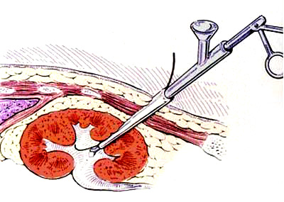
Sometimes if we have a very large stone in the kidney, so called stag horn stone we will do apercutaneous nephrolithotomy. This is keyhole surgery where we puncture the side of the kidney, we go into the kidney, we find the stone, we break the stone either with a lithoclast which is a mini jackhammer or with an ultrasound which breaks up some of the fragments or with a laser and then we remove the fragments that way.
The patients are usually in hospital for two days.
There is some concern with percutaneous nephrolithotomy because we do have to go through the kidney for that procedure but certainly itís preferable to a big open operation.
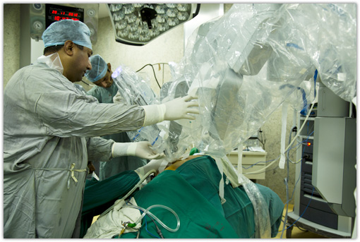
Now occasionally Iíve also removed stones laproscopically with keyhole surgery.
So if we have a very large stone in the kidney, renal, pelvis or ureter we can go in with keyhole surgery and remove it and patients are just in hospital overnight.

The important thing with kidney stones is to get the stones analysed, get blood tests done and to see if patients have abnormal uric acid or calcium level and check that thereís no abnormality.
Sometimes we do a parathyroid level because the parathyroid gland here produces hormones that can lead to increased calcium levels. If patients have recurrent stones I usually refer them to one of my colleagues, one of the renal physicians for a full metabolic evaluation so that we can try to minimise the chance of stone recurrence.
So with kidney stones is to ensure that you do whatever you can to prevent them recurring.
The most important thing is to increase fluid intake, itís very simple, thereís little molecules in the urine and theyíre close together, they join, they form a stone, when theyíre far apart they donít so you have to drink two to three litres a day.
Thereís evidence that home made lemonade is better than water itself.
I tell patients that they should ensure that their urine is clear and theyíre passing urine every two to three hours.
So thatís really what we do with kidney stones.
Itís a common condition but thereís lots of treatment, a lot of good treatments available and if you have recurrent stones you should have a full metabolic evaluation to minimise the chance of them recurring.
UROLOGICAL APPLICATION
DR. VINAY MAHENDRA
MS. Mch (Urol.), DBN (Urol.), FRCS (Glasg.) FRCS (Urol.) Senior Consultant Urologist Laparoscopic And Robotic Urological Surgeon and Renal Transplant Surgeon
ADDRESS:
Apollo Multispeciality Hospitals
58, Canal Circular Road, Kolkata - 700054, West Bengal. India.
FOR APPOINTMENT CALL SECRETARY :
+91 9830216715
(Secretary Amit Roychowdhury : 11:00 A.M. - 4:00 P.M.)
+91 9830492572
(Secretary, Dalia Chatterjee : 11:00 A.M. - 4:00 P.M.)
E-MAIL :
drmahendraappointments@roboticsurgerykolkata.com
WEBSITE :
www.roboticsurgerykolkata.com
POWERED BY :
www.calcuttayellowpages.com